Neonatal Testing
Please record any newborn testing below.
Number of responses: varies. Question type: select one.
|
Yes |
No |
Unsure |
| Head Ultrasound |
28 |
12 |
1 |
| Skull X-Ray |
8 |
22 |
4 |
| Head CT Scan (CAT Scan) |
21 |
14 |
1 |
| Brain MRI |
38 |
4 |
|
| Electroencephalogram (EEG) |
18 |
12 |
6 |
| EKG |
19 |
10 |
4 |
| Renal or Abdominal Ultrasound |
30 |
7 |
2 |
| Hip X-Rays |
14 |
18 |
2 |
| Karotype |
14 |
16 |
9 |
| Microarray |
12 |
16 |
7 |
| FISH |
6 |
16 |
11 |
In regards to the previous question concerning newborn testing, please record any significant and abnormal results.
Number of responses: 22/51. Question type: open-ended.
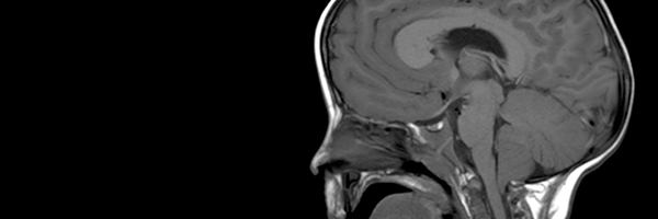
- Head CT and MRI at around 7 months showed enlarged ventricles, abnormal fluid changes, chiari malformation, and macrocephaly.
- Ventriculomegaly and Chiari I Malformation found on MRI.
- MRI showed abnormal brain tissue in the frontal lobe area behind the frontal bossing.
- Ovarian cysts exist on a regular basis (every 3 mos ultrasounds) small but numbers vary and change on each side. No other cysts or concerns to date.
- Brain bleed in the left temporal lobe, hydrocephalus, and white matter missing.
- Somewhat enlarged ventricles on the MRI, normal everything else.
- Enlarged brain ventricles, abnormal white matter, seizure activity, Wolf-Parkinson-White syndrome.
- Enlarged ventricles, and PDA in Heart.
- Mur Mur, thought to have brain damage, collapsed lung, pneumonia.
- Mild pulmonary valve stenosis on EKG, Abnormal white matter on MRI.
- MRI- left hemisphere bigger than right, bleeding in brain (possible delivery trauma) and cyst (spot) in liver (currently being tested).
- Hydrocephaly at 6 months of age.
- Brain MRI: supratentorial ventriculomegaly (greater involvement of occipital horns) and thinning of white matter of the left ventricle. Overall increase in arachnoid spaces. Cyst and cavum septum pellucidum vergae. Altered cortical rutting in left hemisphere perisylvian and rolandic area particularly with areas of polymicrogyria and increased underlying arachnoid space. EEG: abnormal pattern of rhythmic activity based on areas hypersynchronous central front.
- Large ventricles, cyst in brain, oxygen starvation to brain, fluid build up at back of brain. Enlarged kidney due to blockage in urethra.
- Hydrocephaly and hypoplasia Corpus Callosum Hemihypertrophy Epilepsy Hypothyroidism.
- Cranial ultrasounds showed no evidence of hydrocephalus and normal appearances.
- Only Arnold-Chiari I syndrome in Brain MRI. Renal or abdominal ultrasound results were good.
- Ct scan was abnormal, MRI showed polymicrogyria and white matter abnormalities, brainstem atrophy, EEG confirmed seizures.
- Abnormal results with MRI, child was booked in for 3rd ventriculostomy 3 times but blockage corrected itself.
- No hydrocephaly, found fatty filum (operated 2008), one kidney is smaller and no p13 deletion (in question).
|
Were any other newborn tests performed? Please include any abnormal results.
Number of responses: 10/51. Question type: open-ended.
- Eyes were checked for glaucoma.
- Lumbar puncture.
- Oxygen levels were monitored.
- Pelvic X-ray, repeated chest x-rays.
- Audiology tested, results were normal.
- Hearing as premature.
- Test for dwarfism. Ultrasound of heart showed small hole.
- Chromosome tests showed normal male carrier type 46, XY and the 17 hydroxyprogesterone levels were normal.
- ECG & EKG results normal.
- Chest x-ray (found tracheal bronchus that does open to upper-right lung).
|